First and Second Week of Embryonic Development Fertilization Cleavage Implantation Bilaminar Disc Complete Guide 2025
 ---
---
# ⭐ **First & Second Week of Embryonic Development**
*(Fertilization → Implantation → Bilaminar Disc)*
---
## **WEEK 1 — Key Events (Day 0–6)**
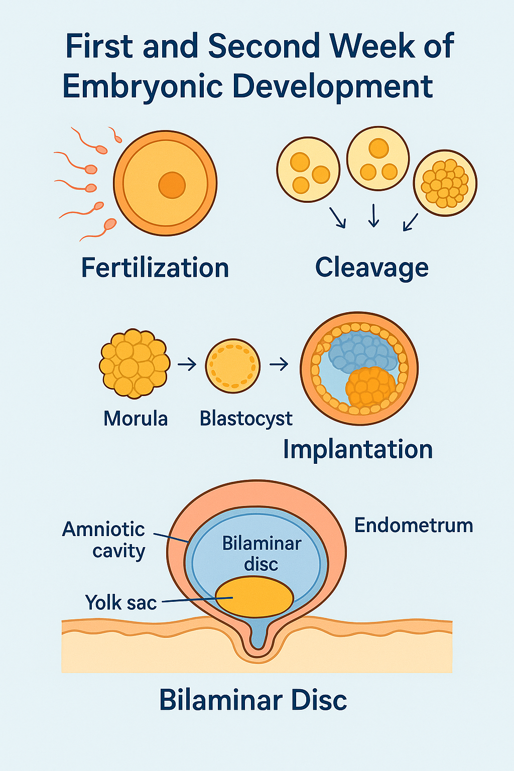
### **1️⃣ Fertilization (Day 0)**
* Occurs in **ampulla of fallopian tube**.
* Sperm penetrates corona radiata → zona pellucida → oocyte plasma membrane.
* Oocyte completes **meiosis II** → forms **zygote (diploid 46,XX / XY)**.
* **Zona reaction** prevents polyspermy.
### **2️⃣ Cleavage (Day 1–3)**
* Rapid mitotic divisions without growth.
* 2-cell → 4-cell → 8-cell stage.
* At **8–16 cells**, forms **morula**.
### **3️⃣ Morula enters uterus (Day 3–4)**
* Surrounded by zona pellucida (prevents ectopic implantation).
### **4️⃣ Blastocyst formation (Day 4–5)**
Blastocyst consists of:
* **Trophoblast → placenta**
* **Inner cell mass (embryoblast) → embryo**
* **Blastocoel** (fluid cavity)
### **5️⃣ Implantation begins (Day 6)**
* Blastocyst attaches to **posterior superior uterine wall**.
* Trophoblast differentiates into:
* **Cytotrophoblast**
* **Syncytiotrophoblast** → invasive, produces **hCG**.
---
## **WEEK 2 — Key Events (Day 7–14)**
**Mnemonic – “Two’s in Week 2”**
* **2 layers** (epiblast, hypoblast)
* **2 cavities** (amniotic cavity, yolk sac)
* **2 trophoblast layers** (cyto + syncytio)
* **2 membranes** (chorion, amnion)
---
### **1️⃣ Completion of Implantation (Day 7–10)**
* Syncytiotrophoblast erodes endometrium → embeds blastocyst fully.
* **Lacunar spaces** form → maternal blood enters → **uteroplacental circulation starts**.
* Trophoblast secretes **hCG**, detectable in maternal blood by **day 8–10**.
---
### **2️⃣ Bilaminar Germ Disc Formation**
Inner cell mass → **two layers**:
* **Epiblast** (forms all 3 germ layers later)
* **Hypoblast** (extraembryonic endoderm for yolk sac)
---
### **3️⃣ Amniotic Cavity Formation**
* Appears above epiblast.
* **Amnioblasts** form **amnion membrane**.
---
### **4️⃣ Yolk Sac Formation**
* Hypoblast cells line blastocoel → **primary yolk sac**.
* Later replaced by **secondary yolk sac**.
---
### **5️⃣ Extraembryonic Mesoderm Development**
* Forms between trophoblast and yolk sac.
* Cavities appear → merge → form **extraembryonic coelom** (→ chorionic cavity).
---
### **6️⃣ Chorion Formation**
Extraembryonic somatic mesoderm + cytotrophoblast + syncytiotrophoblast = **chorion**.
Forms **chorionic villi** → future fetal placenta.
---
### **7️⃣ Prechordal Plate Formation (End of Week 2)**
* Thickened hypoblast area → marks **future head region**.
---
# ⭐ SUMMARY TABLE
| Week | Key Events |
| ---------- | ----------------------------------------------------------------------------------------- |
| **Week 1** | Fertilization → Cleavage → Morula → Blastocyst → Start of implantation |
| **Week 2** | Bilaminar disc → Amniotic cavity → Yolk sac → Chorion → Full implantation → hCG secretion |
---
# ⭐ High-Yield Exam Points (NEET PG / INI CET)
* hCG detectable in maternal blood by **Day 8–10**.
* Syncytiotrophoblast is **invasive** and secretes **hCG**.
* Implantation normally occurs on **posterior superior uterine wall**.
* Embryoblast → **epiblast + hypoblast in week 2**.
* Amniotic cavity from **epiblast**; yolk sac from **hypoblast**.
* Chorion has **3 components**: extraembryonic mesoderm + cytotrophoblast + syncytiotrophoblast.
---HTML Versions